
How a bone actually hardens
Most of the long bones start life as cartilage models. A primary ossification centre appears in the middle of the shaft before birth and turns that cartilage into bone. Secondary centres then appear at each end, in the epiphyses, at fairly predictable ages through childhood.
Between the shaft and each end sits the growth plate, the physis. This is a thin layer of cartilage that keeps producing new tissue, so the bone lengthens. Where a tendon attaches to a bony bump rather than a joint surface, that bump has its own centre, called an apophysis. The tibial tubercle below the knee and the back of the heel are the two apophyses parents hear most about.
Growth ends when the plate stops producing cartilage and the epiphysis fuses to the shaft. Fusion is a process rather than a moment: plates narrow, then close partially, then close fully, usually over 12 to 24 months.
Stage 1
Primary centre (before birth)
Bone replaces cartilage in the middle of the shaft, the diaphysis.
Stage 2
Secondary centres (infancy to childhood)
Separate centres appear in the epiphyses at each end of the bone.
Stage 3
Apophyses appear (roughly 7 to 12 years)
Tendon attachment sites such as the tibial tubercle and calcaneus develop their own centres.
Stage 4
Peak height velocity (girls ~12, boys ~14)
Growth plates are widest, most active and least able to tolerate sudden load increases.
Stage 5
Fusion (mid to late teens)
Plates narrow and close, site by site, ending lengthwise growth at that bone.
The stages every long bone passes through, from cartilage model to fused adult bone.
Labelling the parts of a growing bone
- Diaphysis, the shaft. Site of stress fractures in runners once bone is mature enough to load heavily.
- Physis, the growth plate near each end. Cartilage, weaker than the bone and the tendon around it.
- Epiphysis, the bone end that forms the joint surface. Involved in slipped upper femoral epiphysis and osteochondritis dissecans.
- Apophysis, a bony bump where a tendon pulls. Involved in Osgood-Schlatter (tibial tubercle), Sever's (heel) and Sinding-Larsen-Johansson (lower kneecap).
- Metaphysis, the flared zone between shaft and plate, rich in blood supply and remodelling activity.
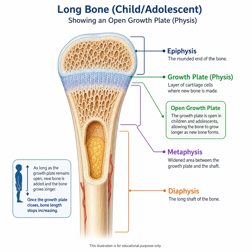
A growing long bone: the growth plate is the cartilage layer between the epiphysis and the shaft.
Typical ossification and fusion ages by site
The figures below are population ranges from skeletal maturity research and radiology references. They are guides for interpreting a pattern, not diagnostic tools, and individual children sit either side of them routinely.
| Site | Centre appears | Fuses, girls | Fuses, boys |
|---|---|---|---|
| Elbow (capitellum, radial head, medial epicondyle) | 1 to 6 years | 13 to 15 years | 15 to 17 years |
| Distal radius (wrist) | 1 to 2 years | 16 to 17 years | 17 to 19 years |
| Iliac crest and pelvis apophyses | 12 to 14 years | 16 to 18 years | 18 to 22 years |
| Femoral head (hip) | Under 1 year | 13 to 16 years | 14 to 18 years |
| Distal femur (knee) | Before birth | 14 to 16 years | 16 to 18 years |
| Tibial tubercle (Osgood-Schlatter site) | 8 to 12 years | 13 to 15 years | 15 to 17 years |
| Lower kneecap pole (Sinding-Larsen site) | 10 to 12 years | 13 to 15 years | 15 to 17 years |
| Calcaneal apophysis (Sever's site) | 5 to 9 years | 13 to 15 years | 14 to 16 years |
| Spine ring apophyses (vertebral endplates) | 9 to 13 years | 18 to 21 years | 18 to 25 years |
Approximate appearance and fusion ages. Girls run earlier at almost every site.
Why girls are roughly two years ahead
Girls enter puberty earlier, so oestrogen rises earlier. Oestrogen drives the growth spurt and then closes the growth plates, which is why girls reach peak height velocity around 11.5 to 12.5 years and finish growing around 15 to 16, while boys peak around 13.5 to 14.5 and can keep growing into their late teens.
The practical consequence in a mixed-age squad is that a 14-year-old girl may have fused knee plates while a 14-year-old boy is at the peak of his spurt with wide-open plates. The same training week lands very differently on those two skeletons.
Approximate age at peak height velocity and at full skeletal maturity.
What the timetable means for injuries
Growth-related conditions are not random. They appear when the relevant apophysis is active and settle once it fuses, which is why Sever's tends to show up a year or two before Osgood-Schlatter, and why heel pain in a 16-year-old boy usually needs a different explanation.
This also explains why a good history includes age, sex, sport and where the athlete is in their growth spurt. Two identical knee complaints in a 12-year-old girl and a 16-year-old boy point at different structures.
| Typical age window | Condition | Structure involved |
|---|---|---|
| 8 to 13 years | Sever's disease | Calcaneal apophysis |
| 10 to 15 years | Osgood-Schlatter | Tibial tubercle apophysis |
| 10 to 14 years | Sinding-Larsen-Johansson | Lower kneecap pole |
| 10 to 16 years | Slipped upper femoral epiphysis | Hip growth plate |
| 11 to 17 years | Growth-related back pain | Vertebral ring apophyses |
| 12 to 17 years | Adolescent patellofemoral pain | Knee joint load tolerance |
| After fusion | Tendinopathy, ACL and ankle ligament injury | Tendon and ligament |
Common conditions matched to the growth stage they belong to.
Using this in training decisions
- Track height every 8 to 12 weeks; a jump of more than about 3 cm in three months signals a fast-growth window.
- During that window, hold total load steady rather than adding volume, and protect jumping and sprinting volume in particular.
- Expect reduced coordination and flexibility during the spurt, and treat it as temporary rather than a technique fault.
- Keep strength work in, well managed resistance training does not damage growth plates and helps the skeleton tolerate sport.
- Group by maturity rather than birth year for collision and high-load sessions where you can.
When to get it checked
See a clinician promptly
Pain that wakes a child at night, a limp lasting more than a few days, hip or groin pain with a limp or restricted rotation, swelling of a joint, a bump that is hot and red, pain after a single significant injury, or any loss of ability to bear weight all need assessment rather than a wait-and-see approach. Growth plate injuries are treatable, but they are best not missed.